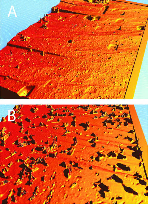
FIG. 2.
(A) Three-dimensional height map of crystallographically defined rhombohedral etch pits in (101̄4) cleavage faces of calcite. (B) False-color image (height map) of a calcite surface with MR-1. The image was taken with a 50× Mirau objective, i.e., the field of view is about 162 by 122 μm, and the height difference is about 1 μm.

