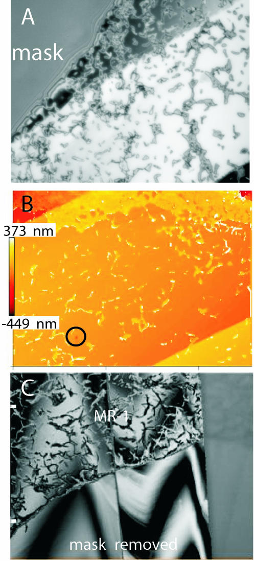
FIG. 4.
(A) Reflected-light image showing MR-1 colonies and single individuals with superimposed interference fringes. (B) Two-dimensional false-color height map calculated from the interferogram. Three different terraces can be identified. At the upper part of the image, MR-1 has started successfully to establish a closed biofilm. (C) Interferogram showing a section of the calcite surface. The upper part of the image is covered with MR-1, while the lower part shows the pristine calcite surface after removal of the mask. No significant height difference was measured between the “reacted” and “pristine” parts of the surface, indicating that MR-1 had successfully blocked the global dissolution of this calcite face.

