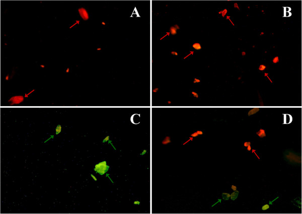
Figure 3.
Photomicrographs of isolated mitochondrial (stained with JC-1 dye) at magnification of 40×. (A) Normal liver, the dye JC-1 concentrates in the matrix and bright red fluorescence was observed; (B) TPW control (400 mg/kg) showed similar features as compared to normal liver; (C) CCl4 treated control liver showed a shift from red to green fluorescence on incubation with JC-1 dye, which indicates damage to the inner mitochondrial membrane (green arrow); (D) TPW (400 mg/kg) + CCl4 treated liver showed red and mild green fluorescence up on incubation with JC-1 dye, which indicates mitochondrial inner membrane integrity was maintained.

