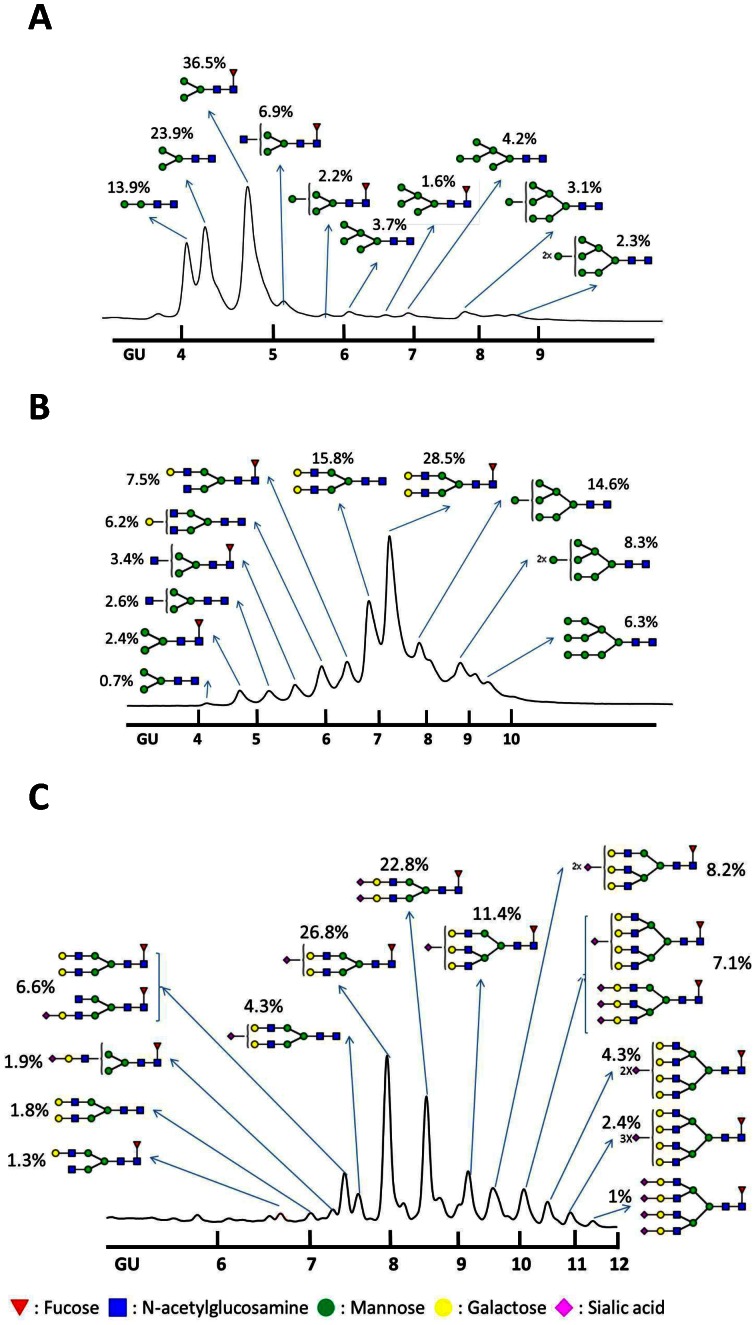
Figure 3. HILIC-HPLC glycan patterns.
Purified recombinant HA proteins were placed on SDS-PAGE gels and deglycosylated using PNGase F. Glycans were isolated from protein backbones and analyzed using HILIC-HPLC to identify individual glycan structures. A dextran ladder was created with HILIC-HPLC and used as a standard to provide glucose unit (GU) values for individual peaks recognized in glycan samples from recombinant HA proteins. Shown are HA N-glycan profiles of (A) Sf9-rHA, (B) Mimic-rHA, and (C) CHO-rHA. Red inverted triangles, fucose; blue squares, N -acetylglucosamine; green circles, mannose; yellow circles, galactose; pink diamonds, sialic acid.

