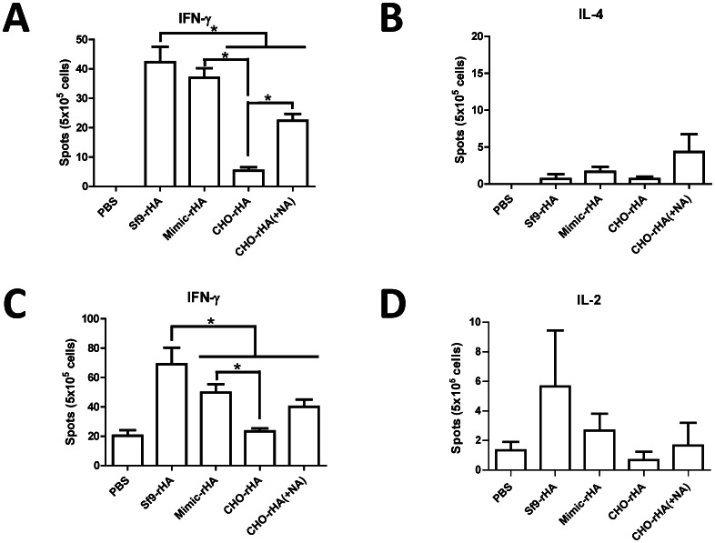
Figure 6. T cell responses in splenocytes and T-cell stimulation by antigen-presenting dendritic cells.
Splenocytes were added to each well in 96-well plates (5×10⁵ cells/well) and stimulated with 1 µg/ml pooled peptides (15-mer overlapping 8 amino acids) spanning the HA1 of H5HA (A/Viet Nam/1203/2004) to determine (A) IFN-γ- and (B) IL-4-secreting T cells using ELISPOT assays. Bone marrow-derived dendritic cells (DCs) were treated with LPS and recombinant HA proteins and then fixed with paraformaldehyde followed by quenching with glycine. Pre-treated DCs were co-incubated with splenocytes from mice immunized with Sf9-rHA for 2 d. Antigen presentation was determined by measuring (C) IFN-γ- and (D) IL-2-secreting T cells using ELISPOT assays. DCs pretreated with LPS and pulsed with PBS were used as a negative control. Data represent mean ± standard deviation. Results were analyzed using two-tailed Student’s t tests with statistical significance at p<0.05 as asterisks indicated.

