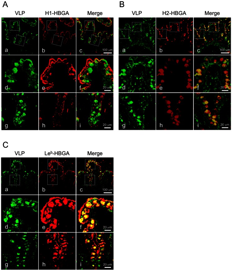
Figure 5. NoV VLPs were colocalized with each HBGA on the surface of epithelial and goblet cells.
Fresh human ileum biopsy specimens from a single individual (individual B) were incubated with 2.5 µg/ml of NoV VLPs in PBS(-) for 1 h at 4°C and subjected to immunofluorescence microscopy. Cryostat sections were incubated with rabbit anti-Ueno 7k VLP serum and mouse anti-type H1, H2 or Leb HBGA antibody and stained with Alexa dye–conjugated secondary antibodies and DAPI. Immunofluorescence images for type H1, H2 or Leb HBGA are shown in panels A, B and C, respectively. Panels d–f and g–i are high magnification views of areas in boxes in panels a–c, respectively. Green, NoV VLPs; red, type H1 HBGA, type H2 HBGA or type Leb HBGA. Scale bars in panel c = 100 µm, and in panel f and i = 20 µm.

