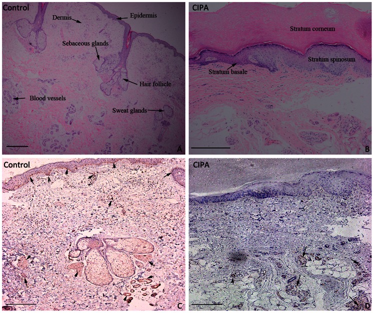
Figure 3. Histopathological examination of a skin biopsy.
(A) Normal epidermis and appendages in healthy control samples (H&E staining). (B) Hyperkeratosis, acanthosis and irregular stratum basale, few sweat glands, hair follicles and sebaceous glands in the CIPA patient's samples (H&E staining). (C, D) NSE immunoreactivity stains of control and CIPA patient's sections. Arrow indicates the area with positive staining. (Immunohistochemical staining, Bar = 100 µm)

