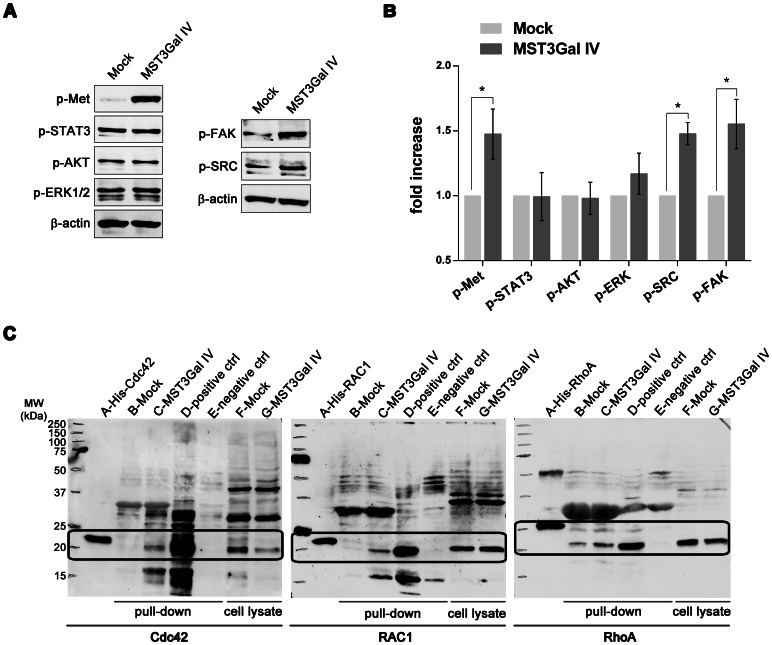
Figure 5. Evaluation of downstream effectors of c-Met activation.
A – Increased levels of p-FAK and p-Src proteins in MST3Gal IV cells. The contribution of other effector proteins, such as AKT, ERK, FAK and Src was evaluated by Western blot for their phosphorylated forms in Mock and MST3Gal IV cell lines, and expression of β-actin protein was used as protein loading control. Results show increased levels of phophorylated FAK and Src supporting their involvement as downstream effectors of phosphorylated c-Met (p-Met). B – Analysis of 5 independent Western blot of c-Met, STAT3, AKT, ERK, FAK and Src phosphorylated forms in MOCK and MST3 Gal IV cells showing statistically significant increased levels of p-FAK and p-Src, concomitantly with increased phosphorylated c-Met. Results are presented as means ± SD. C - Evaluation of Cdc42, Rac1 and RhoA GTPases as potential modulators of c-Met activation by pull-down assay of their activated forms. Western blot analysis of pull-down proteins evidence an increased activation of Cdc42, Rac1 and RhoA in MST3Gal IV cell line. A-GTPase WB protein positive control (His-Cdc42, His-Rac1 and His-RhoA); B-Mock total cell protein pull down; C-MST3Gal IV total cell protein pull down; D-Mock total cell protein pull down with previous GTPases activation (pull down positive control); E-Mock total cell protein pull down with previous GTPases inhibitors (pull down negative control); F-Mock total cell protein input; G-MST3Gal IV total cell protein input. Highlighted areas represent regions of interest regarding the specific protein migration.

