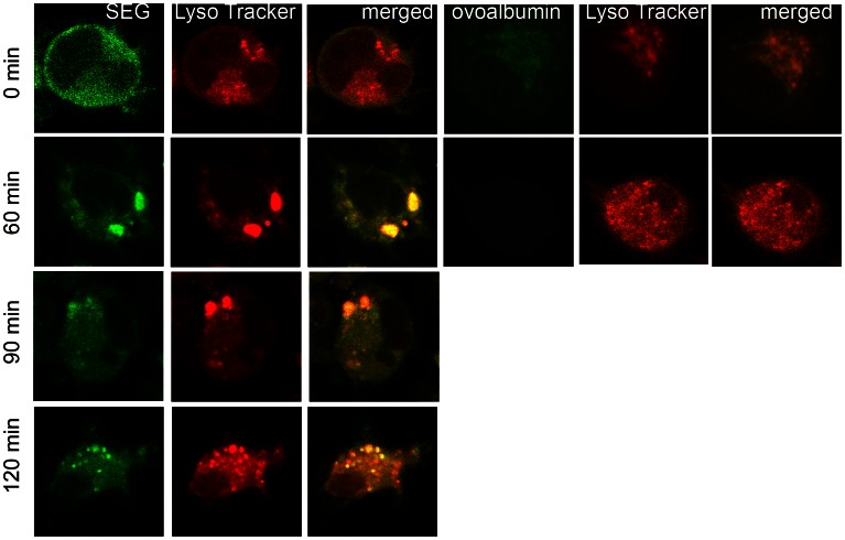
Figure 2. After incorporation in DCs, SEG co-localize with LysoTracker in acidic organelles.
DCs were cultured on poly-L-lysine treated slides, pulsed 1 h at 37°C with 50 µg/ml SEG-FITC and 50 µM Lysotracker RED (Molecular Probes, Invitrogen), washed and cultured further for different time periods (0–120 min). Fluorescently labeled cells were fixed with paraformaldehyde at 4°C, and cover slides mounted using anti-fade mounting media (Vector Laboratories). Image capture was performed using a Nikon C1 confocal laser scanning microscope with a PlanApo 60× Oil AN1.40 lens. For detection of FITC a 490 nm laser was used. A 546-nm excitation wavelength and a 590-nm emission wavelength were used for LysoTracker. Analysis by confocal microscope (6000×) shows that SEG barely co-localized at acidic compartment at time point 0, but clearly co-localized with LysoTracker at the 60 min time point and later. Cells pulsed with 50 µg/ml OVA-FITC for 1 h showed co-localization of OVA with Lysotracker at time point 0, but 60 min later only LysoTracker remained inside the cell. Figure shows a representative experiment of 3.

