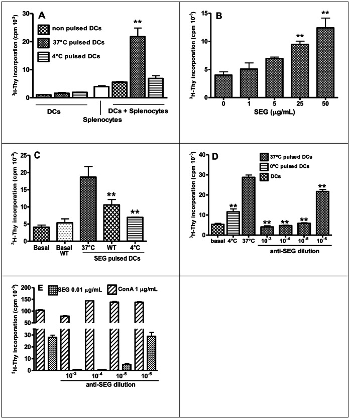
Figure 6. DCs induce superantigenic response after SEG uptake.
DCs were pulsed 1 h at 37°C or 4°C with SEG, washed, and co-cultured with isogenic splenocytes in triplicate; controls of DCs with no addition of splenocytes were conducted in parallel. (A) 3H-Thy incorporation for DCs, splenocytes, and DCs plus splenocytes. (B) DCs were pulsed with different concentrations of SEG for a dose response curve, and co-cultured with splenocytes in triplicates. (C) Splenocyte proliferation induced by SEG-pulsed DCs and SEG-pulsed DCs in the presence of Wortmannin, performed in triplicates. (D) Rabbit anti-SEG serum was added to the cultures of pulsed DCs and splenocytes to specifically block SEG function. (E) Splenocytes were cultured in the presence of 0.01 µg/ml SEG or 1 µg/ml ConA, with the addition, or not, of different dilutions of rabbit anti-SEG serum. One representative experiment is shown of 3 to 5 experiments. **p<0.01.

