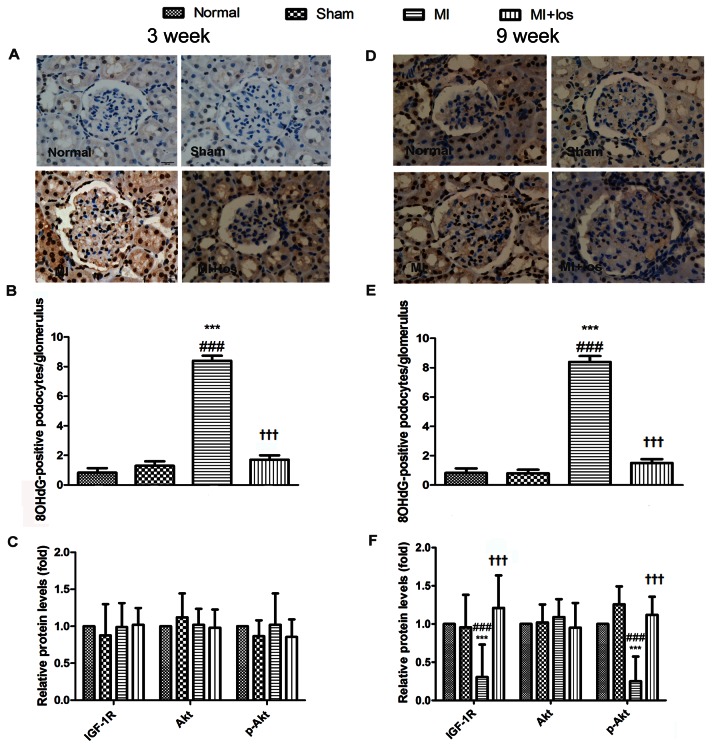
Figure 4. Potential mechanisms involved in podocyte injury in normal, sham, MI and MI+los rats.
(A) Representative immunohistochemical staining for 8-OHdG (original magnification, ×400) at 3 weeks. (B) The average number of 8-OHdG-positive podocytes per glomerulus at 3 weeks. (C) Expressions of IGF-1R, Akt and phosphorylated(p)-Akt proteins at 3 weeks. (D) Representative immunohistochemical staining for 8-OHdG (original magnification, ×400) at 9 weeks. (E) The average number of 8-OHdG-positive podocytes per glomerulus at 9 weeks. (F) Expressions of IGF-1R, Akt and phosphorylated(p)-Akt proteins at 9 weeks. Data are presented as the mean ± SEM. ###P < 0.008 vs. normal; ***P < 0.008 vs. sham; ††† P < 0.008 vs. MI. MI, myocardial infarction; los, losartan; 8-OhdG, 8-hydroxy-2'-deoxyguanosine; IGF-1(R), insulin-like growth factor-1(receptor).

