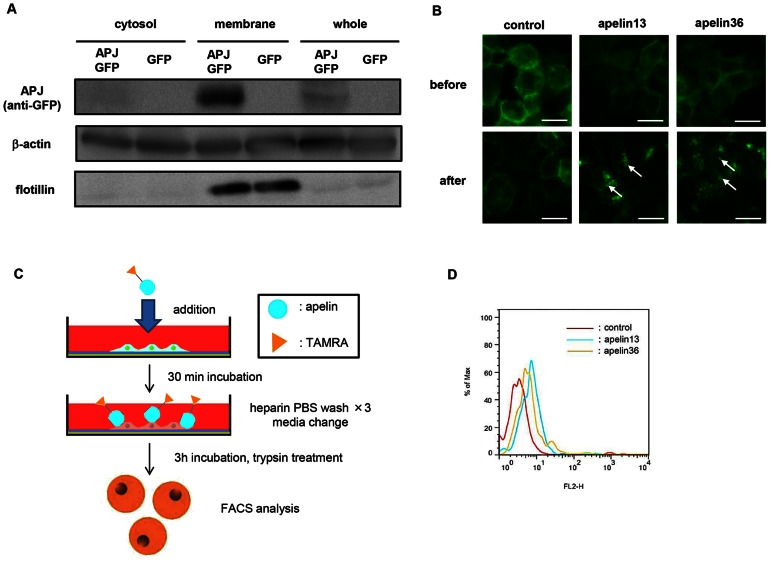
Figure 1. Internalization of apelin via cell surface APJ.
(A) Western blot analysis of mouse APJ-GFP fusion protein on NIH-3T3 cells. Anti-GFP antibody was used to detect APJ-GFP. Flotillin is the positive control for membrane proteins on NIH-3T3 cells. β-actin was used as the internal control. (B) Internalization of mouse APJ-GFP with apelin. NIH3T3-APJ-EGFP cells were incubated in the presence or absence of apelin 13 or apelin 36. White arrows indicate internalized APJ. Bar indicates 20 µm. (C) Scheme for apelin internalization analysis. NIH3T3-APJ-EGFP cells were incubated with apelin (13 or 36)-TAMRA for 3 hrs, washed with heparin PBS to remove non-specifically bound apelin-TAMRA from the cell membrane. Cell membrane proteins were trypsinized and cells were analyzed by flow cytometry. (D) APJ-mediated uptake of apelin-TAMRAs by flow cytometry.

