
Figure 5. Phenotype designation reveals differences in gray matter and white matter density.
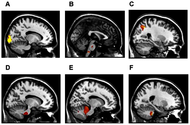
(A) START subjects (in contrast to controls) had less gray matter volume in the left lingual gyrus extending into the left cuneus (P<0.025) and (B) right pons and right medulla (P<0.02) (C) STOPP subjects (in contrast to controls) demonstrated a trend of less gray matter in the right superior parietal lobule extending into the right precuneus (P<0.07). (D) START subjects had reduced white matter volume (in contrast to STOPP) in the left pons (P<0.004) and (E) left cerebellar tonsil and left pyramis (P<0.012) (F) Analyses also demonstrated START subjects (in contrast to STOPP) had decreased gray matter in the right culmen extending into the right fastigial and left dentate nucleus of the cerebellum (P<0.035). All P values are corrected for age, gender and multiple comparisons using non-stationary cluster correction.