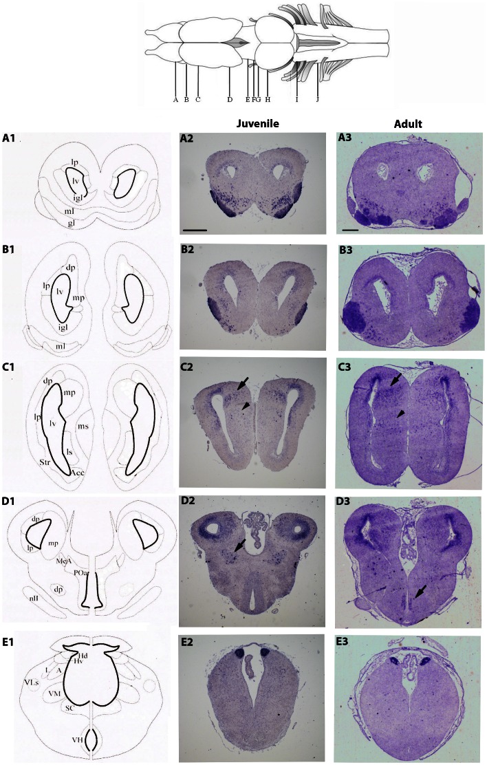
Figure 1. Expression pattern of NeuroD1 in the juvenile (A2–E2) and adult (A3–E3) X. laevis brains.
A1–E1) Schematic coronal illustrations of the corresponding transverse sections of a juvenile X. laevis brain (NF stage 66). The drawing at the top of the figure shows a dorsal view of the X. laevis brain. The letters correspond to the rostro-caudal location of sections as depicted in the whole brain drawing. Arrows and arrowheads in C2, C3, D2, and D3 highlight less conspicuous areas of labeling. Abbreviations are defined in Table 1. The anatomical drawings are from [55], with modifications of basal ganglia subdivisions according to [56]. For all images, dorsal is to the top. Scale bar = 400 µm in A2–E2, and 100 µm in A3–E3.

