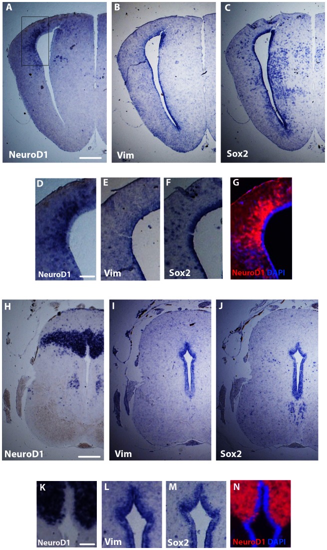
Figure 3. Expression patterns of NeuroD1, Sox2 and Vimentin in the juvenile X. laevis brain.
In situ hybridizations on coronal sections of cerebral hemispheres (A–G) and cerebellum (H–N). In cerebral illustrations, D, E and F are high magnifications of A, B and C, respectively. In cerebellum illustrations, K, L and M are high magnifications of H, I and J, respectively. To allow merge with the DAPI staining, colors of high magnification illustrations D and K were negatively inverted in photos G and N, respectively. For all images, dorsal is to the top. Scale bar = 220 µm in A–C and H–J; 95 µm in D–G and K–N.

