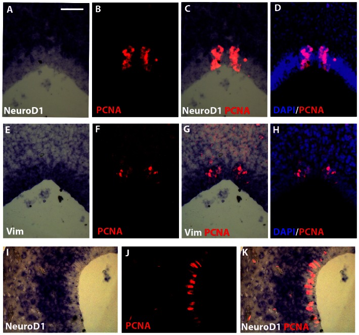
Figure 4. NeuroD1/PCNA and Vimentin/PCNA double stainings in the cerebellar and pallial regions of juvenile X. laevis brain.
Coronal sections at the level of cerebellum (A–H) or pallium (I–K). In situ hybridization using a NeuroD1 (A–D and I–K) or a Vimentin (E–H) probe combined with PCNA immunohistochemistry. For all images, dorsal is to the top. Scale bar = 30 µm.

