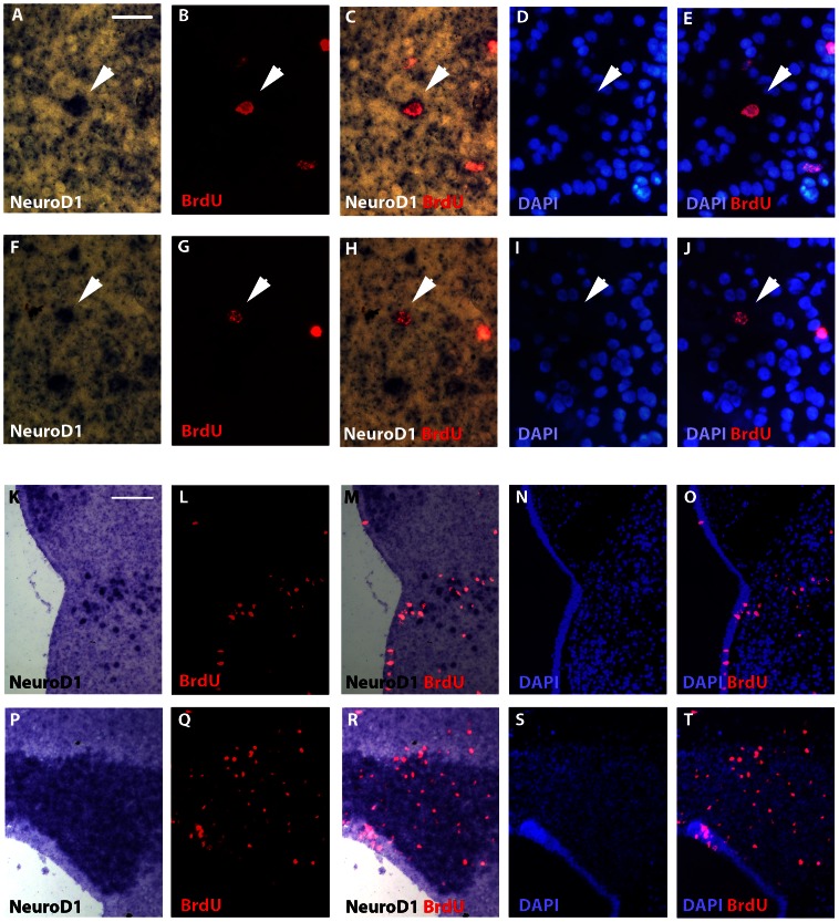
Figure 5. NeuroD1/BrdU double stainings on telencephalic and cerebellar sections of a juvenile X. laevis brain.
(A–J) Telencephalon high magnifications of NeuroD1 in situ hybridizations (A, C, F and H) combined with BrdU immunodetections (B, C, F and H) after 14-days BrdU post-administration time. Arrows indicate double stained cells. DAPI stainings are indicated to certify the presence of the nucleus. (K–T) Low magnifications of the above NeuroD1/BrdU/DAPI triple labelling experiments showing larger view of telencephalon (K–O) and cerebellum (P–T). For all images, dorsal is to the top. Scale bar = 15 µm in A–J; 45 µm in K–T.

