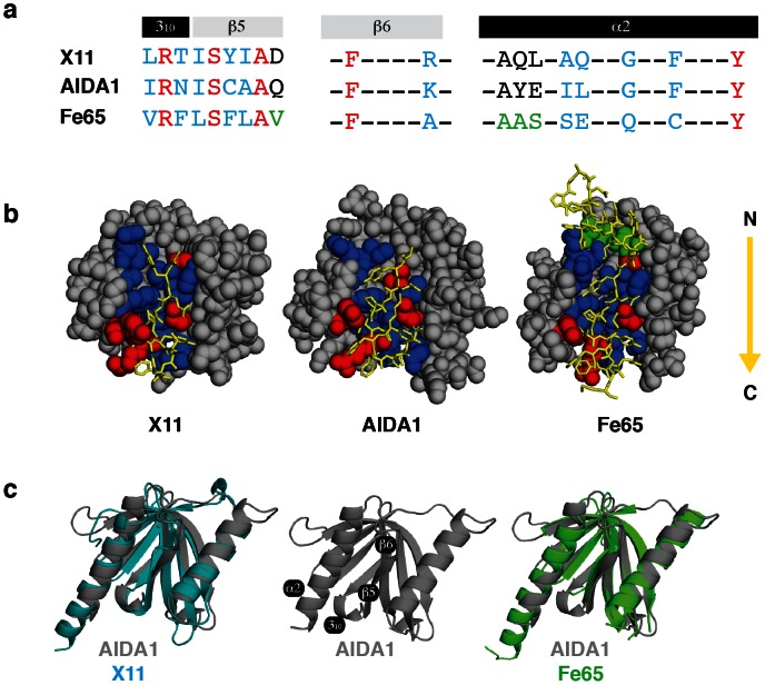
Figure 3. Interaction of a APP derived peptide (GYENPTYKFFE, shared among all) with the X11, Fe65 and AIDA1 PTB domains.
(a) Sequence alignment of amino acids that contribute to the binding cleft; identity in red, homology in blue. The Fe65 PTB domain recognized a longer APP sequence, amino acids that extend its cleft are shown in green. (b) APP (yellow, in stick format, N-C direction follows the arrow) interacting with the X11/Fe65 as determined from their respective X-ray structures and with AIDA1 determined from a molecular docking simulation. (c) Backbone alignment of the AIDA1 (grey), X11 (cyan) and Fe65 (green) PTB domain in the same orientation as (b) with the binding cleft facing forward.

