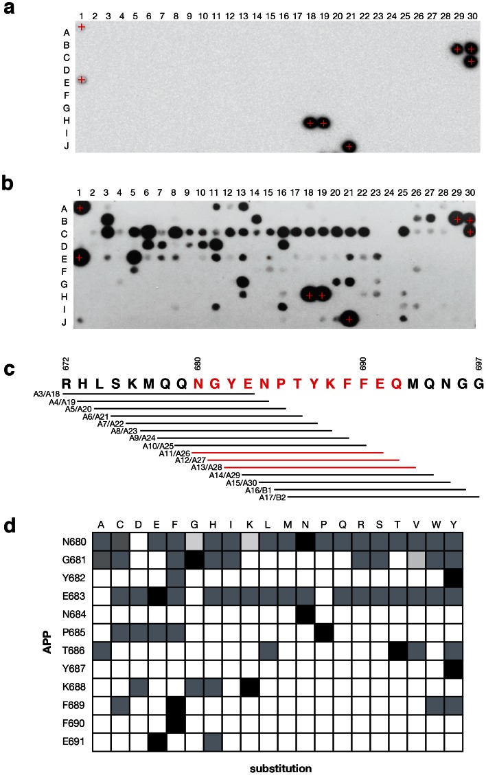
Figure 4. Amino acid preferences of the AIDA1 PTB domain for APP determined from a peptide array.
A list of peptides on the array are provided in supplementary material. (a) The array probed with anti 6xHis mAb only. Positive control 6xHis peptides are identified by a +. (b) The array probed with 6xHis-AIDA1 PTB domain. (c) Sliding window peptide scan of 12-mers spanning aa. 672–697 of APP. Peptides are duplicated on the array; for example, at A3 and A18. Since peptide content per spot can vary, if a signal was observed at the exposure presented it was deemed to be interaction. (d) Results of a window scan across the APP C-terminal sequence and an exhaustive positional scan. Grey boxes indicate binding was observed, regardless of signal intensity.

