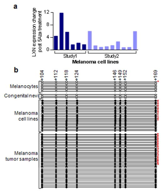
Figure 3. Promoter region hypermethylation leads to silencing of LXN in melanoma.
(a) Analysis of microarray data from two previous experiments showing restoration of LXN expression in melanoma cell lines upon treatment with methylation reversing drug 5 Aza 2 deoxycytidine. Study1: light red (Muthusamy et al 2006); Study2: dark red (unpublished data). (b) Sanger bisulfite sequencing of the LXN gene promoter CpG island in melanocytes, melanoma cell lines and tumor samples. CpG positions are indicated by circles in scale to their location in the promoter region. Clear circles indicate absence of methylation, filled circles represent methylated cytosine. The numbers at the top indicate distance from the transcription start site. Stars indicate that the methylation status of these samples were described previously (Muthusamy et al 2006)

