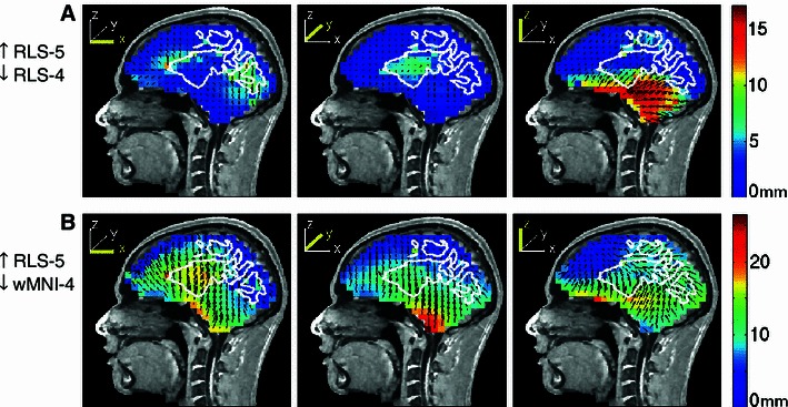
Fig. 11.

Equivalent dipole source localization error directions (arrows) and magnitudes (colors) relative to simulated dipole projections using a four-layer reference head model (S1) for EEG data simulated using a five-layer BEM head model including a white matter layer. The white matter boundary in the five-layer model is outlined in white. Other details as in Fig. 3
