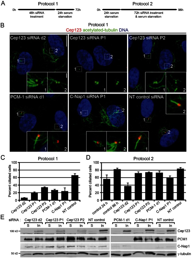
Fig. 4. Cep123 is required for primary cilium formation but not maintenance.
(A) A schematic explaining the two protocols used to determine if Cep123, PCM-1 or C-Nap1 is required for the formation (protocol 1) or maintenance (protocol 2) of a primary cilium. (B) Immunofluorescent images of RPE1 cells stained with DAPI to label the DNA (blue) and antibodies against acetylated tubulin (green) and Cep123 (red). Scale bars: 10 µm and inset 1 µm. (C,D) Quantification of the number of ciliated cells, where the results represent the means of three independent experiments (n = 200–450), with the s.d. shown. The depletion of Cep123, PCM-1 or C-Nap1 prevented primary cilium formation, resulting in a significant decrease in the number of ciliated cells (student's t-test: Cep123 siRNA d2, P = 1.8×10⁻⁵; Cep123 P1, P = 6.0×10⁻⁵; Cep123 P2, P = 2.5×10⁻⁴; PCM-1, P = 1.5×10⁻⁴; and C-Nap1, P = 1.7×10⁻³). However, Cep123 and PCM-1 appeared to be dispensable for primary cilium maintenance while C-Nap1 was only partially required (student's t-test: Cep123 siRNA d2, P = 7.9×10⁻⁴; Cep123 P1, P = 1.2×10⁻²; Cep123 P2, P = 7.2×10⁻³; PCM-1, P = 2.3×10⁻²; and C-Nap1, P = 2.3×10⁻³). (E) Western blotting of soluble and insoluble extracts prepared from siRNA-treated RPE1 cells demonstrated the depletion of the appropriate protein.

