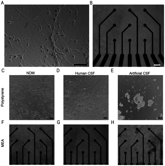
Fig. 1.

Neuronal cells grew well on (A) laminin-coated polystyrene and (B) PEI and laminin-coated MEA plates in NDM before the artificial or human CSF exposure. Two weeks after the artificial or human CSF exposure, neuronal cells grew in NDM (C,F) and human CSF (D,G) but detached in artificial CSF cultures (E,H). Scale bars: 100 µm.
