Fig. 4.

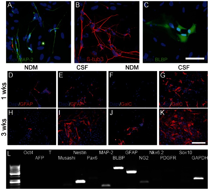
Representative pictures of MAP-2- and β-tubulin3-positive neuronal cells (A,B) and BLBP-positive radial glial cells (C) in CSF cultures. Cells cultured in NDM expressed glial proteins for astrocytes (GFAP) but not for oligodendrocytes (GalC) at first week's time point (D,F) and only a few glial cells could be detected at third week (H,J). In comparison, both GFAP- and GalC-positive glial cells were detected in CSF cultures already at first week (E,G) and the amount of glial cells increased towards the third week (I,K). Cells in CSF cultures expressed oligodendrocytical genes NG2, Nkx6.2, PDGFR, and Sox10 in addition to other neural markers after 4 weeks of differentiation (L). Scale bars: 50 µm in A–C; 100 µm in D–K.
