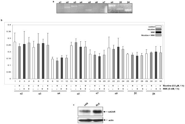
Figure 1.
Expression of nAChR in rat lung epithelial RLE and murine lung cancer LKR cells. a. Total RNAs from the cells were isolated. Equal amount of RNAs was reverse-transcribed, and the expressions of nAChR subunits were identified by PCR. b. The cells were treated with nicotine (0.5 μM) or NNK (1 μM), or treated with nicotine for 30 min prior to adding NNK for 1 h, total RNAs were extracted. After reverse-transcription, real-time PCR was performed. The abundance of nAChRs was normalized to actin. Error bars represent the standard deviation over 3 independent experiments. c. Expression of nAChR in RLE and LKR cells.

