
Figure 1. The CB is a dynamic nuclear domain responsive to many stimuli.
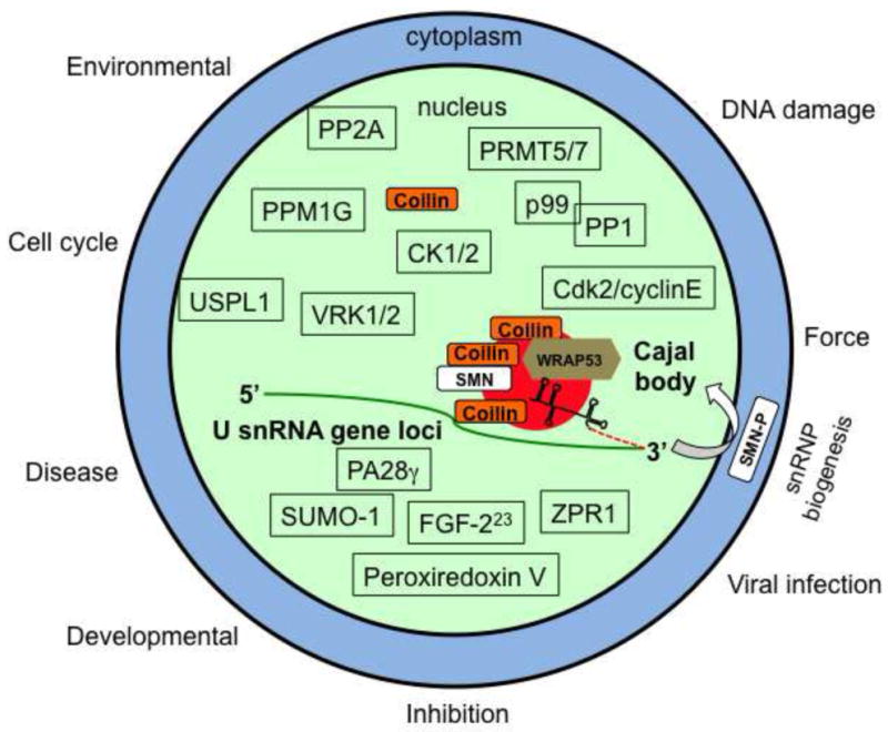
Major structural components of the CB include coilin, WRAP53, SMN and snRNPs. Since CBs are known to associate with many gene loci, including those giving rise to U snRNA, such as U2 snRNA, it is possible that the CB participates in the initial 3′ end processing event of the nascent pre-U snRNA (stem loop structure with dashed red line). Mature snRNPs, scaRNAs and TERC are not shown. Besides accumulations in the CB, every known protein component of the CB also resides elsewhere in the cell. The majority of coilin, for example, is nucleoplasmic. Shown above the CB are enzymes known or suspected to modify coilin or other components of the CB: PPM1G (protein phosphatase, Mg2+/Mn2+ dependent, 1G) (Hearst et al., 2009, Petri, Grimmler, 2007), CK1/2 (casein kinase 1 (our unpublished results) and 2) (Hebert and Matera, 2000), VRK1/2 (vaccinia-related kinase 1 and 2) (Sanz-Garcia et al., 2011), PRMT5/7 (protein arginine methyltrasferase 5 and 7) (Boisvert, Cote, 2002, Gonsalvez, Tian, 2007), protein phosphatases PP2A and PP1(γ), with adaptor subunit p99 (PNUTS) (Kitao et al., 2008, Lyon et al., 1997, Moorhead et al., 2007, Renvoise et al., 2012) and Cdk2/cyclinE (Liu et al., 2000). The ubiquitin-specific protease-like 1 (USPL1) SUMO isopeptidase protein has been shown to impact coilin localization (Schulz, Chachami, 2012). Shown below the CB are signaling molecules that impact or are found in CBs: PA28γ (proteasome activator) (Cioce et al., 2006), FGF-223 (fibroblast growth factor) (Bruns et al., 2009), ZPR1 (zinc finger protein 259) (Gangwani et al., 2001), SUMO-1 (Navascues, Bengoechea, 2008) and Peroxiredoxin V (Kropotov et al., 2004). The biogenesis of U1, U2, U4 and U5 snRNPs require cytoplasmic processing steps under the control of SMN. Cytoplasmic SMN is hyperphosphorylated (SMN-P) relative to that found in the nucleus, due in part to the action of the nuclear phosphatase PPM1G (Grimmler, Bauer, 2005, Petri, Grimmler, 2007). Arrayed along the outside of the cell are stimuli that affect CB assembly.