
Figure 4.
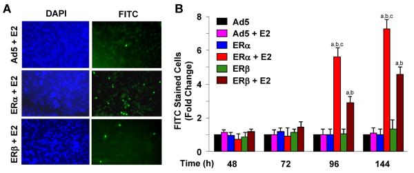
Fig. 4 ERs in the presence of E2 induce apoptosis of MDA-MB-231 cells. Cells were infected with recombinant adenoviruses as a function of time in the absence or presence of 10−9 M E2. Infected cells were subjected to a TUNEL assay. a Representative TUNEL images at 96 h post-infection are shown. FITC indicates apoptotic cells that incorporated the FITC-conjugated dUTP into the fragmented DNA. DAPI was used to stain cell nuclei. b Graph, which is the mean ± SEM of three independent experiments, depicts the number of cells stained with FITC at different time points. Superscript (a) denotes responses that are significantly different from those observed with cells infected with the parent Ad5 in the absence of E2. Superscript (b) indicates responses that are significantly different from responses to ERs in the absence of E2 at the corresponding time point. Superscript (c) denotes responses that are significantly different from responses to ERβ + E2