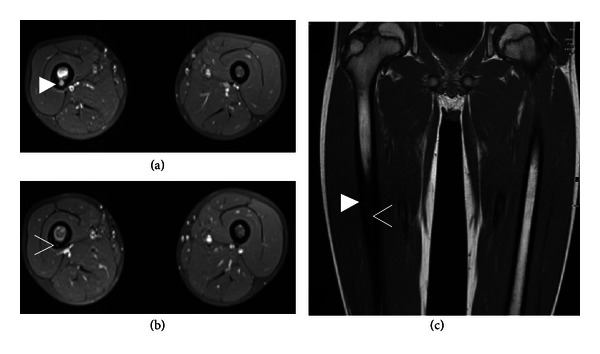
Figure 2.

Contrast-enhanced MRI reveals a multicentric osteoid osteoma with two nidi in the femur diaphysis with a larger nidus of 6 × 5 mm (solid arrowhead in (a) and (c)) and a smaller 2 × 2 mm nidus inferiorly (open arrowhead in (b) and (c)). Note the thickened cortical bone and the bone marrow edema of the right femoral diaphysis. (a) and (b) contrast-enhanced T1 fs MRI; (c) T1 TSE MRI.
