Fig. 7.
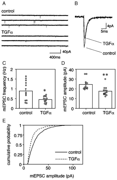
TGFα decreases mEPSC amplitudes in multipolar neurons. Cortical cultures were treated with or without TGFα for 5 days from 8 DIV and assayed at 13–14 DIV. (A) Spontaneous mEPSCs were recorded from multipolar neurons by a whole-cell patch-clamp technique. (B) Thirty mEPSC responses were calculated from multipolar neurons and averaged. (C) mEPSC frequencies were analyzed for multipolar neurons of control culture or TGFa-treated culture (n = 12 each, *P < 0.05, U = 32, Mann–Whitney U test). (D) mEPSC amplitudes of multipolar neurons were plotted (n = 12 each, **P < 0.01, U = 18, Mann–Whitney U test). Average quantal amplitudes of 100 events for the neurons that were grown with or without TGFa. (E) Cumulative amplitude histograms of mEPSCs were plotted for TGFa-treated and control multipolar neurons (P < 0.0001, Kormogorov–Smirnov test).

