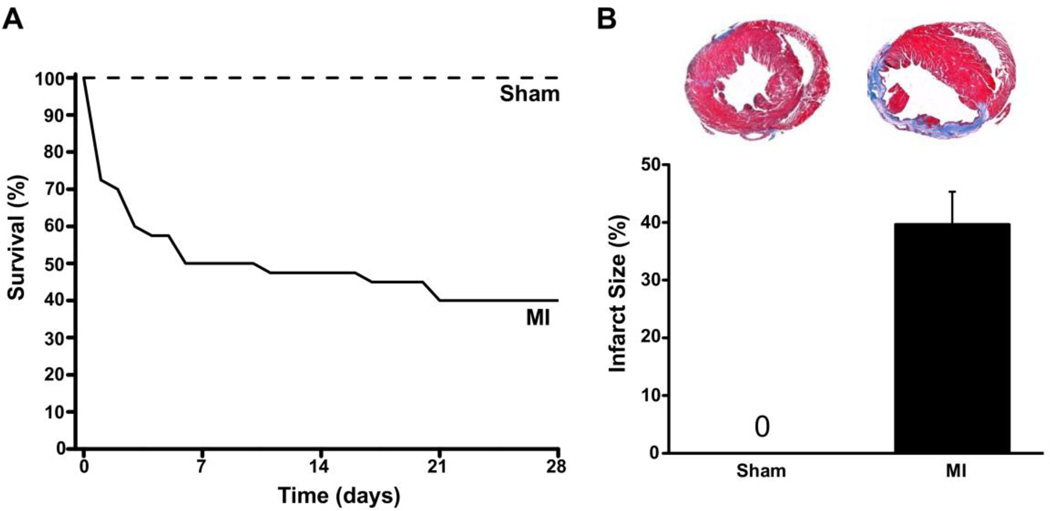
Fig. 1.
Generation of chronic MI mouse model. Compared to sham mice (n=10), the survival of mice after permanent ligation of the mid-LAD was significantly reduced, with a survival rate of only 40% after four weeks (A). Panel B shows the average infarct size of the mice that survived to the 4 week time point (n=16). Insets: representative cross-sections stained with Trichrome-blue from sham and post MI hearts. The infarct scar (blue stain) occupies the LV anterior wall and the large cavity area indicates chamber dilation.

