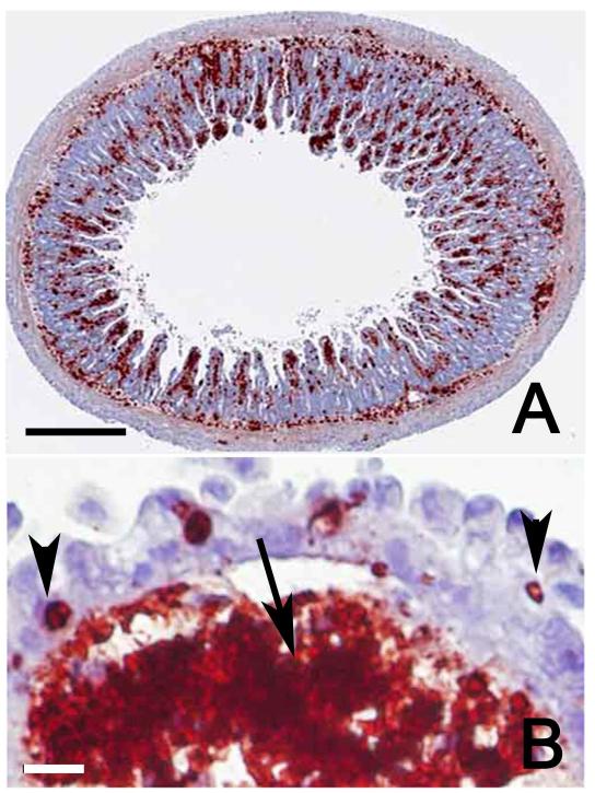
Figure 1.
Section of ileum of a Swiss Webster mouse, 96 hr after feeding VEG strain oocysts. Note extensive parasitisation of the lamina propria. All red stained bodies are tachyzoites. Immunohistochemical staining with rabbit T. gondii polyclonal antibodies. A. Low magnification showing cross section. Bar=250 μm. B. Higher magnification of a villus showing numerous tachyzoites and spilled antigen (arrow) in the lamina propria. A few tachyzoites (arrow heads) are present in enterocytes but the surface epithelium is intact. Bar=25 μm.

