
Figure 1.
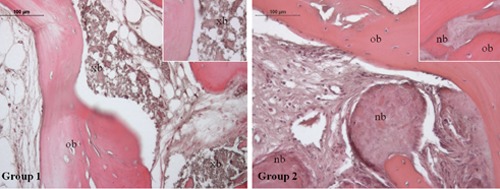
Hematoxylin and eosin staining of Group 1 and Group 2 specimens. Magnification 20×. Group 1: bone tissue specimens obtained from equine-derived bone substitute grafted area; Group 2: bone tissue specimens obtained from calvaria bone grafted area. Inset (40x) shows, in group 1 specimens, grafted biomaterial particles, in group 2 specimens, large mineralized areas within which newly formed bone can be recognized because of osteocyte lacunae lack and absence of lamellar organization; xb, xenogenous bone; ob, old bone; nb, new bone.