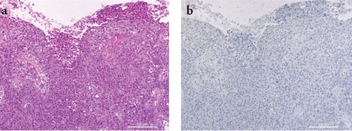
Figure 5.

Representative staining results in a case of squamous cell carcinoma. a) H&E staining showing a squamous cell ulcerated tumor composed of nests of atypical epithelial cells extending into the dermis; b) IMP-3 staining was negative. Scale bars: 200 µm.
