Figure 1.

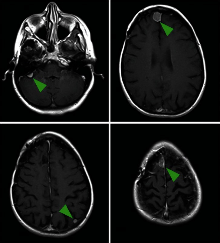
Axial images of a gadolinium-enhanced magnetic resonance imaging of the brain. The patient had a symptomatic right frontal meningioma treated with surgery, but had four additional separate meningiomas in discrete locations

Axial images of a gadolinium-enhanced magnetic resonance imaging of the brain. The patient had a symptomatic right frontal meningioma treated with surgery, but had four additional separate meningiomas in discrete locations