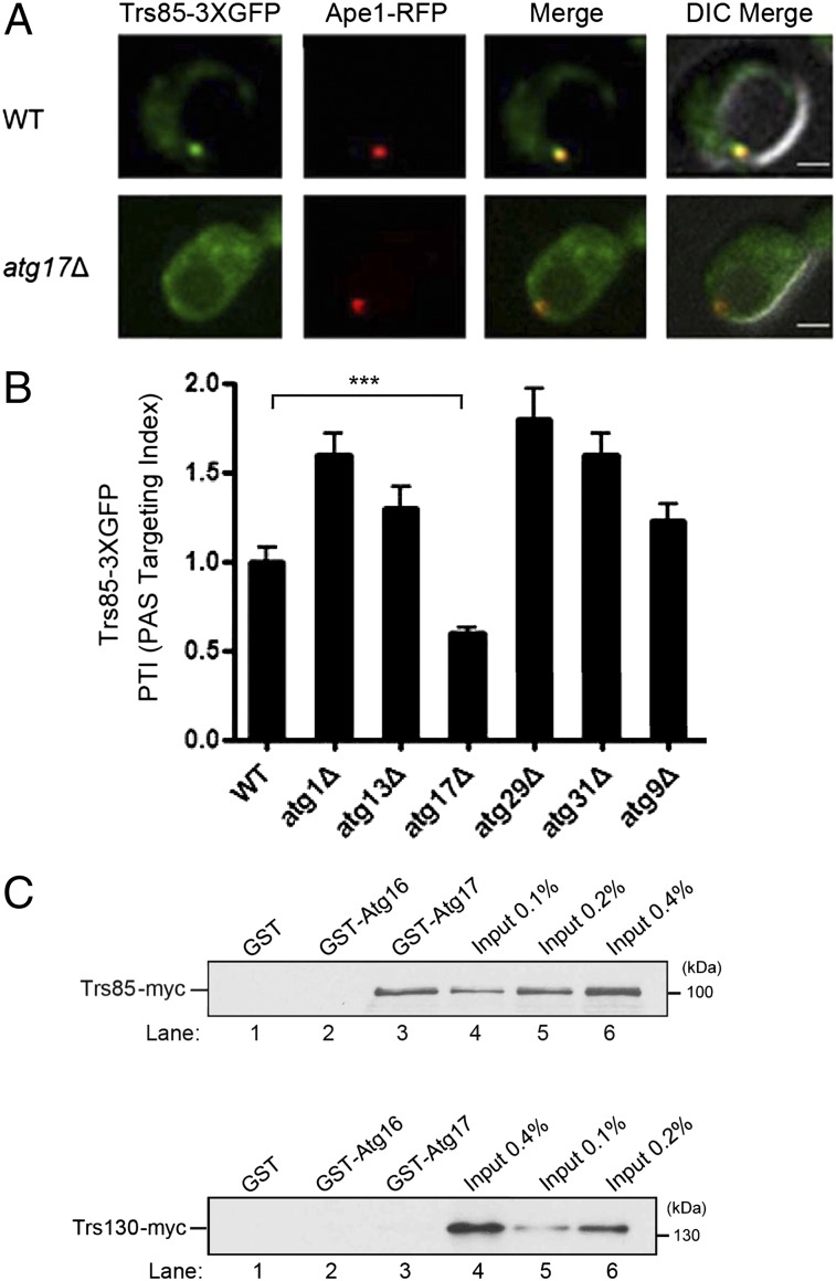
Fig. 2.
The localization of Trs85–3XGFP to the PAS is disrupted in the atg17Δ mutant. (A) Cells expressing Trs85–3XGFP and Ape1–RFP were grown to log phase in synthetic complete (SC)–Leu medium, pelleted, and resuspended in SD-N medium for 4 h. (Scale bar, 2 μm.) (B) Quantitation of the data in A, and several other atg mutants, in our laboratory strain background, which is derived from S288C. The PAS targeting index (PTI) was calculated in 50 cells by multiplying the percent of cells that contain colocalized Trs85–3XGFP and Ape1–RFP to the total Trs85–3XGFP signal that resides at the PAS. The PTI in wild-type was set at 1.00. Error bars represent SEM, n = 150 cells from three separate experiments. ***P < 0.001 Student t test. (C) Trs85 specifically binds to Atg17. Yeast lysate containing Trs85–myc (Top) or Trs130–myc (Bottom) was incubated with glutathione-Sepharose beads. Bound protein was eluted and analyzed.

