Fig. 2.
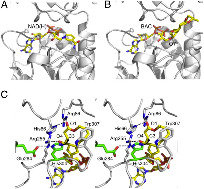
Model of BAC binding to BARS. View of the BARS nucleotide binding site with (A) the NAD(H) cofactor bound (PDB ID code 1HKU) and (B) the modeled BAC molecule. Position 7 of BFA, where the conjugation between ADPR and BFA takes place to form BAC, is indicated. (C) Stereoview of the BAC binding site. Residues relevant to BAC interaction and to catalysis are shown in stick representation (white and green, respectively) and labeled. Hydrogen bonds are indicated as dashed lines, whereas the nucleophilic attack between His304 and the C3 atom of BAC is indicated by an arrow. Hydrogen atoms are not shown.

