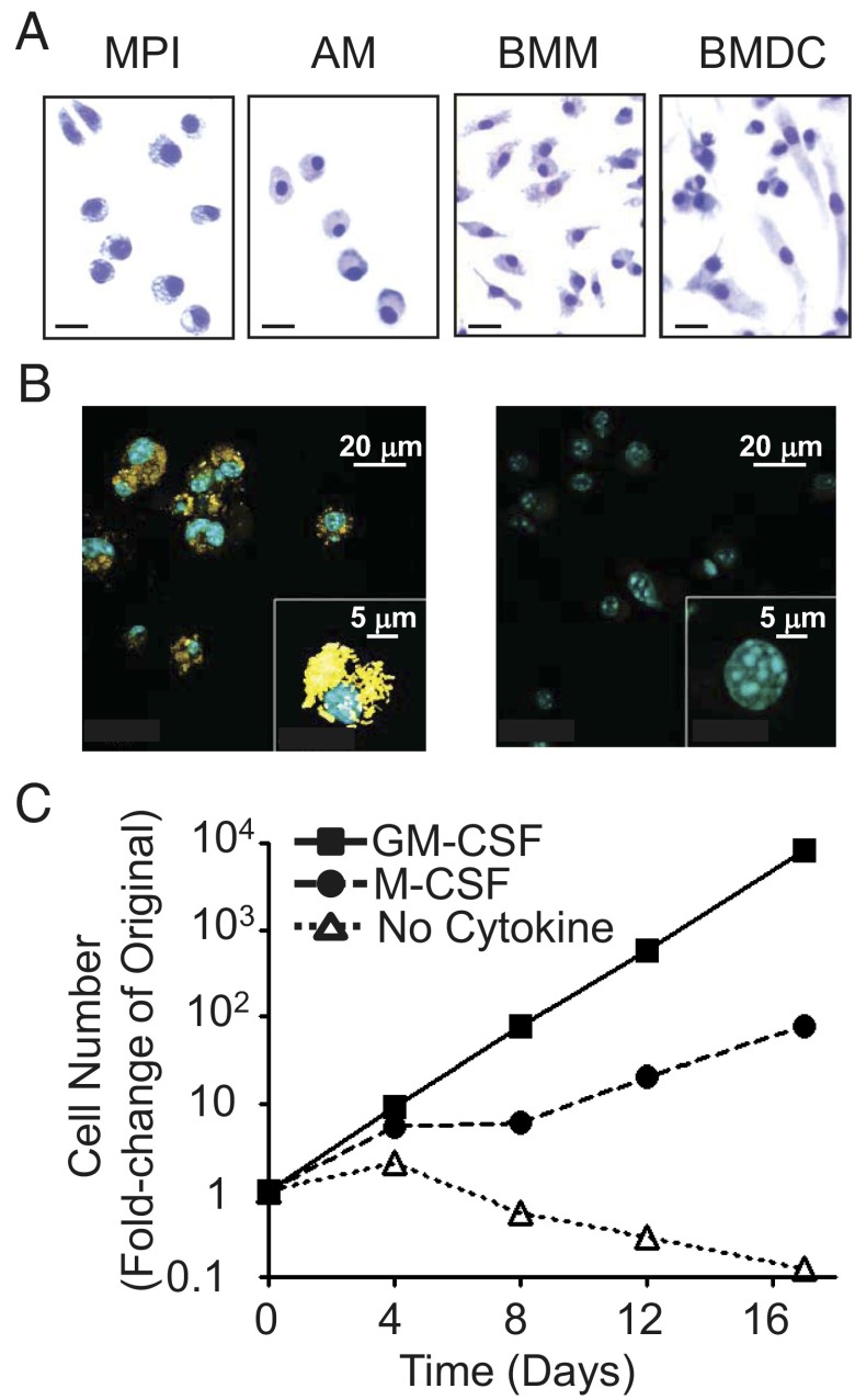
Fig. 1.
MPI cells are factor-dependent, self-renewing phagocytes. (A) Giemsa staining of MPI cells, AMs, BMMs, and BMDCs. (Scale bars, 20 μm.) (B) Phagocytosis of Alexa 647-stained P. acnes (Left, yellow) in MPI cells and in mock-treated cells (Right) using confocal microscopy. Blue, DAPI-stained nuclei. (Insets) Single P. acnes- and mock-treated cells at high power. (C) Growth curve of MPI cells with GM-CSF (30 ng/mL), M-CSF (30 ng/mL), or without any growth factor.

