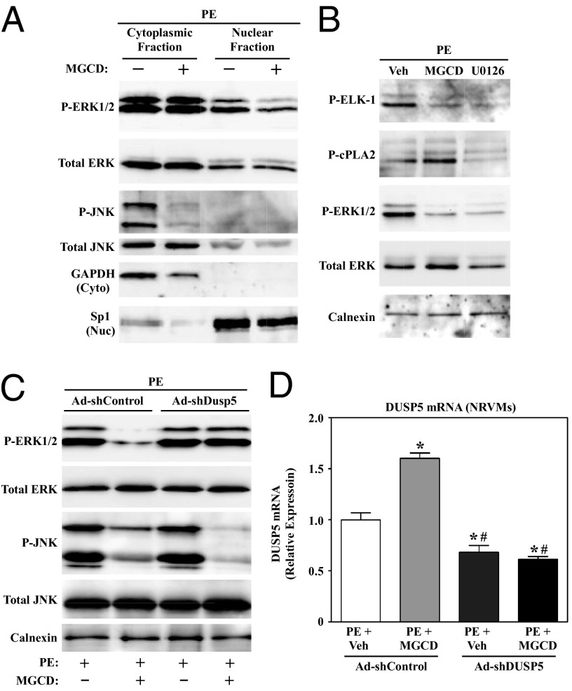
Fig. 4.
Class I HDAC inhibition selectively blocks nuclear ERK1/2 signaling in cardiomyocytes via induction of DUSP5. (A) NRVMs were treated with PE for 48 h in the absence or presence of MGCD0103. Nuclear and cytoplasmic protein fractions were prepared and immunoblotted with the indicated antibodies. GAPDH and Sp1 served as controls to establish the purity of cytoplasmic and nuclear fractions, respectively. (B) NRVMs were left untreated or treated for 48 h with PE in the absence or presence of MGCD0103 or the MEK inhibitor, U0126. Immunoblot analysis was performed with whole-cell lysates to assess the degree of phosphorylation of a cytoplasmic ERK1/2 substrate (cPLA2) and a nuclear ERK substrate (ELK-1). (C) NRVMs were infected with adenoviruses encoding shRNA to knockdown expression of endogenous DUSP5 (Ad-shDUSP5) or scrambled negative control (Ad-shControl); multiplicity of infection (MOI) = 50 for each virus. After 24 h of infection, cells were treated with PE in the absence or presence of MGCD0103 for an additional 48 h and whole-cell lysates were analyzed by immunoblotting. (D) RNA was harvested from parallel plates of NRVMs to assess DUSP5 mRNA expression by qPCR. n = 3 plates of cells per condition. *P < 0.05 vs. PE plus vehicle in cells infected with Ad-shControl; #P < 0.05 vs. PE plus MGCD0103-treated cells infected with Ad-shControl.

