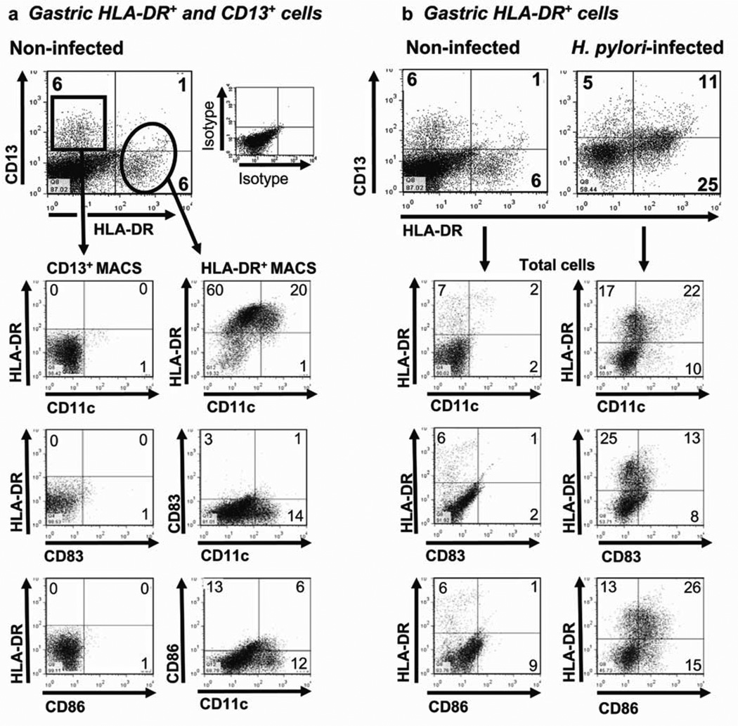
Figure 1.
Phenotype of gastric mucosal cells from non-infected and H. pylori -infected subjects. (a) CD13+ and HLA-DR+ cells isolated from the gastric mucosa of a representative noninfected subject and enriched by magnetic antibody cell sorting (MACS) separation (n =10). (b) Phenotypic characterization of HLA-DR+ DCs among cells isolated from a representative, noninfected subject (n =4) and an H. pylori -infected subject (n =4). Numbers in the quadrants indicate percentage of cells in the respective quadrant.

