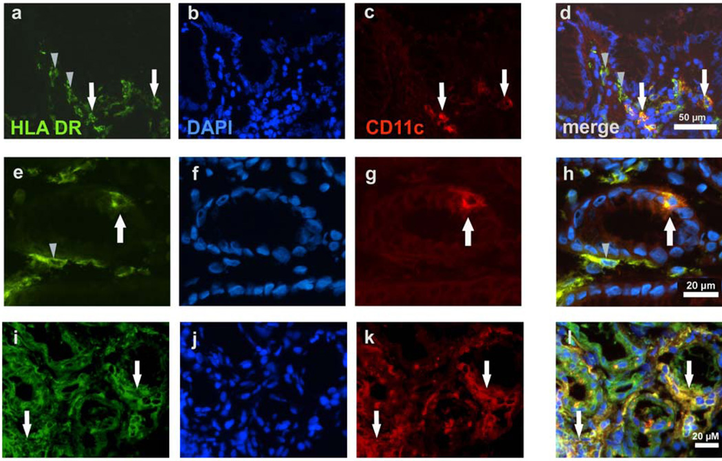
Figure 2.
Immunohistochemical identification of dendritic cells (DCs) in the gastric mucosa of noninfected and H. pylori -infected subjects. (a-d) Gastric mucosa from a noninfected subject contains HLA-DR+ cells in which a small proportion are HLADR+/ CD11c+. (e-h) Gastric mucosa from a noninfected subject shows gastric glands with an intraepithelial HLA-DR+/CD11c+ cell. (i-l) Gastric mucosa from an H. pylori infected subject contains many HLA-DR+ cells in which a large proportion are HLADR+/ CD11c+. Note the increased HLA-DR expression in gastric epithelial cells in the infected subject (i-l) compared with epithelial cells in the noninfected subject (a-h). (a,e,i) HLA-DR-FITC; (b,f,j) 46-diamidino-2-phenyl indole (DAPI) nuclear stain; (c,g,k) CD11c-TXRD (red); (d,h,l) merge. White arrows indicate HLA-DR+/CD11c+ DCs; gray arrowheads indicate HLA-DR+/CD11c− cells, likely immature DCs. Results are representative of four noninfected and four H. pylori -infected subjects.

