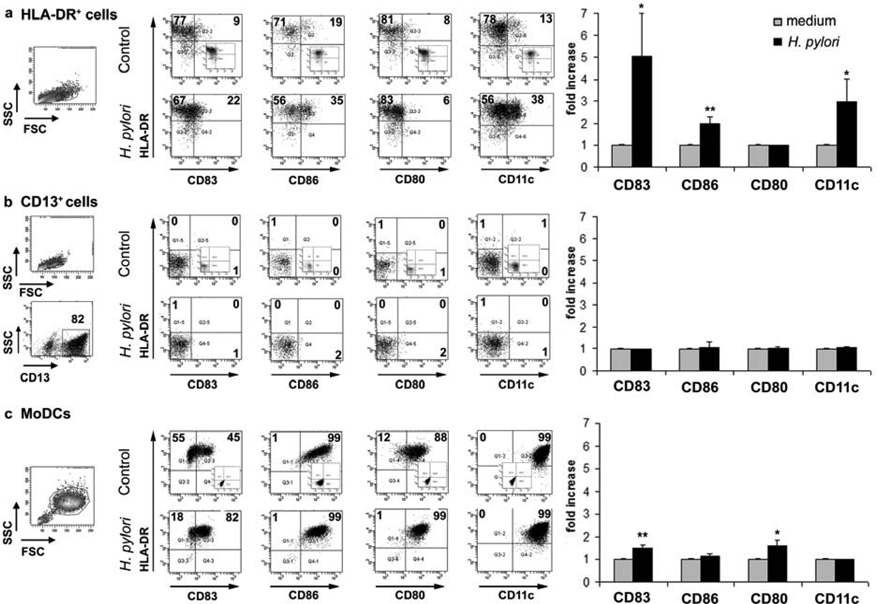
Figure 3.
Maturation of gastric HLA-DR+ dendritic cells (DCs) and monocyte-derived DCs (MoDCs) in response to H. pylori. (a) HLA-DR+ DCs and (b) CD13+ cells isolated from the gastric mucosa of an H. pylori -negative donor, and (c) MoDCs generated from H. pylori -negative donor monocytes were exposed to live H. pylori (strain 60190, multiplicity of infection 20) or medium alone (control) for 15 h, followed by fluorescenceactivated cell sorting (FACS) analysis. Numbers in dot plots indicate percentage of cells in respective quadrants. Insets show isotype-matched controls. Bar graphs on the right show mean fold increase ± s.e.m. (n =3) in the expression of the indicated markers, calculated as f(x) = % stained cells (sample)/% stained cells (medium control); * P ≤0.05, ** P ≤0.01; unpaired Student’s t -test.

