
Figure 4.
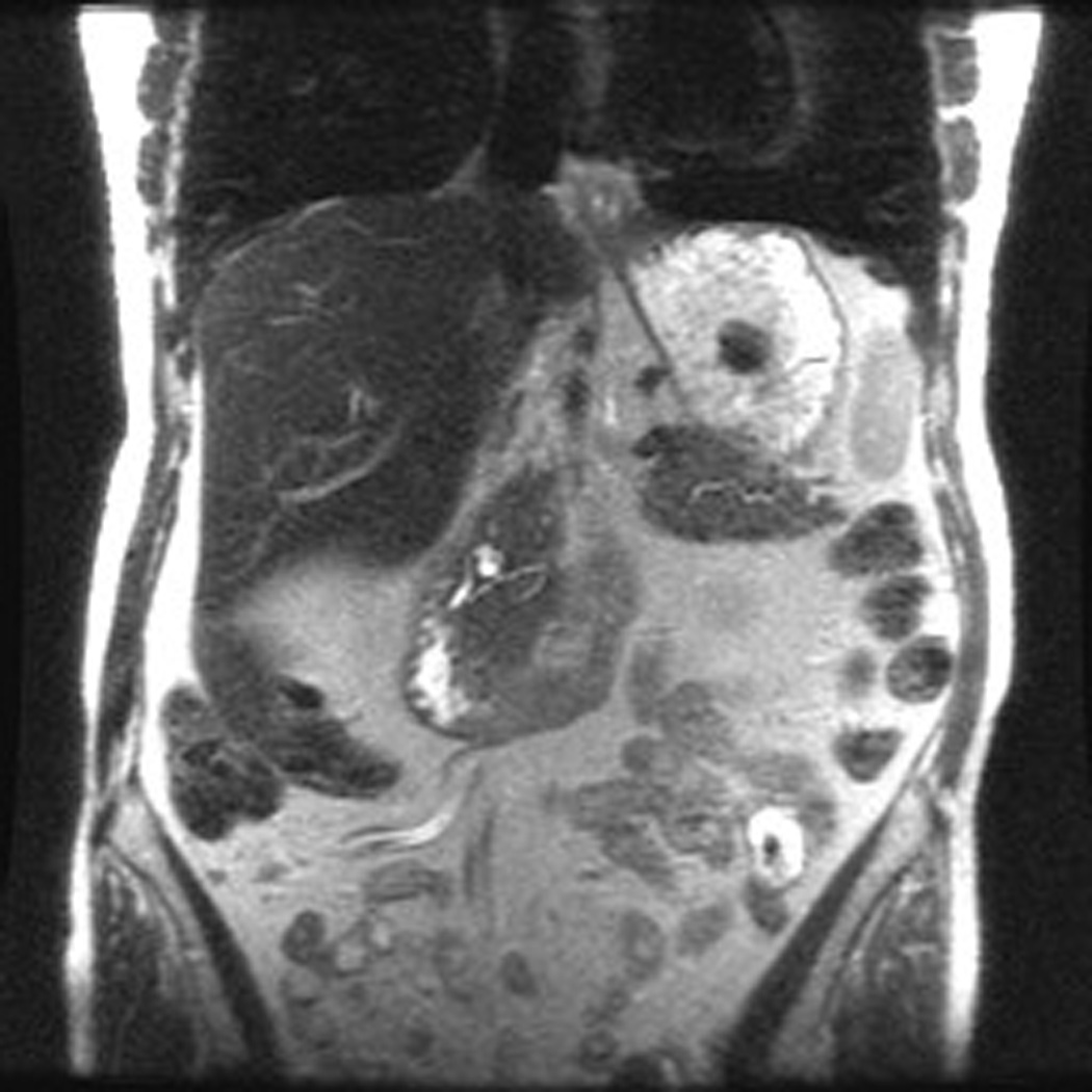
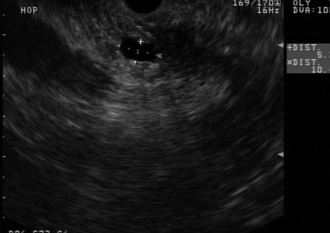

Patient 1 is a 73 year old woman with one FDR and one SDR (father diagnosed at age 70 and paternal grandmother at age 77). On initial MRCP, patient 1 was found to have a cystic pancreatic head lesion. Subsequent EUS showed two cystic lesions in the head and neck of the pancreas, communicating with branches of the PD. FNA of cyst fluid was performed, cytology from which was suspicious for malignant cells. Based on this, surgical resection was recommended, and the patient underwent a pylorus sparing pancreaticoduodenectomy. Pathology revealed multifocal IPMN of borderline malignant potential, predominately involving side branches. She recovered well from surgery and continues with surveillance of her remnant pancreas.