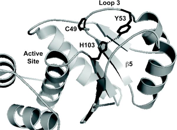
Figure 3. Y53-H103 Hydrogen Bonding in PRL-1.
PDB code 1XM2 was used to illustrate the proximity of the Y53 and H103 side chains.36 In the figure, the side chain of these residues, as well as C49 are shown as sticks. Various important regions of the protein, including loop 3 (L3) and the central β-strand (β5) are labeled. In the oxidized crystal structure (PDB 1ZCK),32 the interaction between Y53 and H103 is perturbed as these side chains move away from one another. This figure was generated using Pymol.

