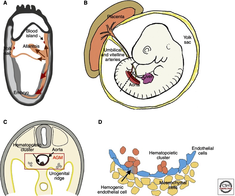
Figure 1.
Ontogeny of the mouse hematopoietic system. (A) During gastrulation of the mouse conceptus, emerging mesodermal cells (brown arrows) migrate to the extraembryonic compartments (yolk sac and allantois). Blood islands begin to form in the yolk sac upon interactions of the mesoderm (orange) with the endoderm (gray). (Red arrows) Mesoderm migrating into the embryo proper. (B) Embryonic day 10.5 (E10.5) mouse embryo. Hematopoietic sites include the aorta (AGM region), yolk sac, umbilical and vitelline arteries, placenta, and liver. (Dotted line) The transverse section in panel C. (C) Transverse section showing the AGM region of an E10.5 mouse embryo. Urogenital ridges are lateral to the aorta. A hematopoietic cluster on the ventral wall of the aorta is shown. (D) Close-up of the aorta showing a hemogenic endothelial cell transitioning to a hematopoietic cluster cell. Nonhemogenic endothelial cells (blue) and mesenchymal cell (yellow) are shown.

