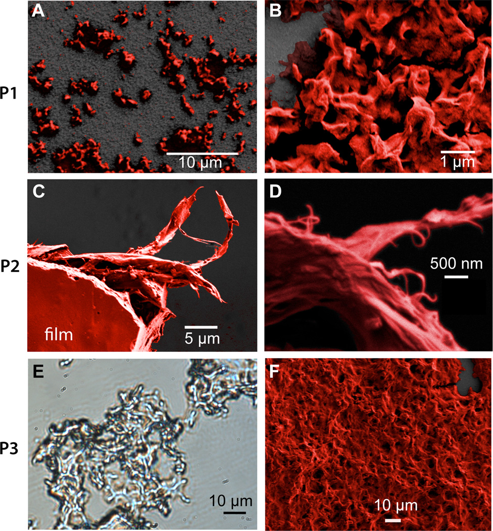
Figure 6.
Electron and optical microscopy. (a. & b.) SEM micrographs of P1 showing absence of film formation. (c. & d.) SEM micrograph of P2 showing a piece of polymer film that tore apart during sample preparation leaving behind fibrous shreds at two magnifications. (e.) phase contrast light micrograph of P3 showing complex morphology. (f.) SEM micrograph of P3 showing an amorphous morphology. SEM Images have been colorized and the dark gray back-ground is the substrate.

