Figure 1.

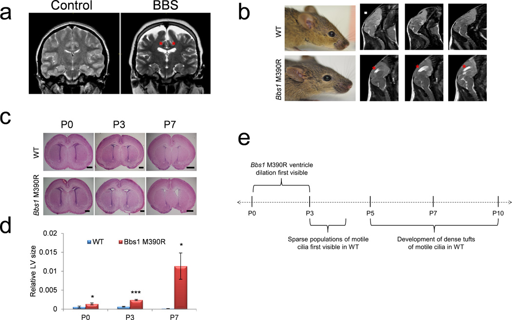
Hydrocephalus in BBS mutant mice occurs before motile cilia develop. (a) T2-weighted coronal MRIs of a Bardet-Biedl syndrome patient and age and sex matched control showing ventriculomegaly of the lateral ventricles (red asterisks). (b) Left, picture of 3 month old WT and Bbs1M390R/M390R mice showing a normal cranial vault in WT and Bbs1M390R/M390R. Right, T2-weighted sagittal MRIs showing hydrocephalus of the lateral ventricles (red asterisks) in a Bbs1M390R/M390R mouse. (c) Histology of WT and Bbs1M390R/M390R neonates showing perinatal onset of hydrocephalus in mutant pups and (d) the quantitations showing ventricular dilation at P3 and P7. (e) Timeline of the genesis of hydrocephalus in BBS mutant mice relative to motile cilia development showing that Bbs1M390R/M390R mice develop hydrocephalus prior to the development of motile cilia13,14. All error bars represent means ± s.e.m. *P<0.05, ***P<0.0005, results from unpaired t tests. All experiments utilized at least 3 mice per group and genotype. Scale bars equal 1 mm (b) and 500 µm (P0 and P3) and 1 mm (P7, c). E, embryonic day; LV, lateral ventricle; P, postnatal day.
