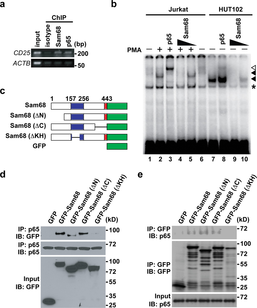
Figure 3. Sam68 is an integral component in the NF-κB complex binding to the CD25 κB site.
(a) HUT102 cells were used for ChIP assays with indicated antibodies. The recruitment of indicated proteins to the κB region of CD25 promoter or ACTB promoter in the immunoprecipitates was detected by PCR. (b) Nuclear extracts of HUT102 cells (lanes 7–10) and Jurkat cells (lanes 1–6) treated with (+) or without (−) PMA (50 ng/ml, 30 min) were analyzed by EMSA with 32P-labeled CD25 κB oligonucleotides. Supershift analysis was conducted with p65 or different doses of Sam68 antibodies (lanes 3–5 and 8–10). CD25 κB DNA binding complexes in Jurkat cells and HUT102 cells are labeled with filled triangles, and the supershifted bands and nonspecific bands are labeled with an open triangle and an asterisk, respectively. (c) Schematic diagram of the full-length and truncated mutants (ΔN lacks residues 1–102, ΔC lacks residues 347–443, and ΔKH lacks residues 165–224) of Sam68 fused with EGFP. The hnRNP K homology (KH) domain (residues 157–256) and nuclear localization signal (NLS) are labeled in blue and red, respectively. (d–e) Whole cell lysates from HEK293T cells transfected with GFP vehicle or indicated GFP-fused full-length or truncated Sam68, were immunoblotted (IB) directly or after immunoprecipitation (IP) with p65 antibody (d) or GFP antibody (e) for indicated proteins.

