Figure 1.

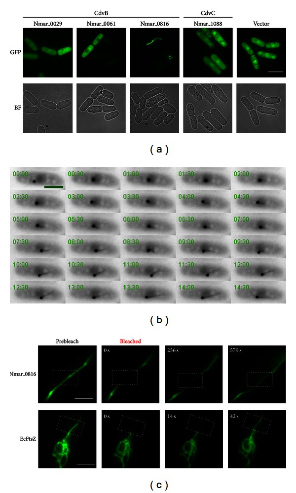
N. maritimus CdvB (Nmar_0816) forms filament-like structures in yeast. (a) Images of fission yeast cells expressing N. maritimus CdvB paralogs and CdvC fused with GFP. BF: bright field; Scale bar: 10 μm. (b) Time-lapse images of Nmar_0816 polymer formation in fission yeast. Cells carrying pREP42-Nmar_0816-GFP were cultured in the absence of thiamine for 24 h at 24°C and monitored for GFP signals. Frames were captured at 15 s intervals. (Video S1). Scale bar: 5 μm. (c) Time-lapse images of the Nmar_0816-GFP polymers versus the E. coli FtsZ-GFP polymers upon fluorescence recovery after photobleaching (FRAP). Dotted rectangle indicates bleached region. Scale bar: 3 μm.
