FIG. 1.
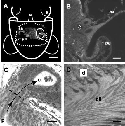
V. fischeri must travel down narrow, ciliated ducts to colonize the light organ of E. scolopes. (A) Schematic of a ventral view of a hatchling squid superimposed over a confocal micrograph of the light organ (e = eye). The light organ is circumscribed by the posterior portion of the funnel (dotted white lines) (modified from reference 31). The pores (*) leading to the ducts of the organ are located at the base of a pair of “appendages” (aa = anterior appendage; pa = posterior appendage), components of two complex ciliated fields, one on each lateral surface of the organ (circled in white), that facilitate colonization by symbionts. Bar, 250 μm. (B) Higher-magnification confocal image of one side of the organ in the region of the ducts. The most medial duct (⋄) was used for all analyses in the present study. Bar, 25 μm. (C) Histological section of an uninfected juvenile light organ. Each pore (p) is an opening to a ciliated, microvillous duct that leads (arrows) to one of three epithelium-lined crypts (c) on each side of the light organ. In the crypts, the bacteria interact with the epithelium and a population of macrophage-like hemocytes, which appear in this image as central in a crypt space. The dashed line indicates the position in the duct at which all confocal analyses were performed. Bar, 25 μm. (D) Transmission electron micrograph demonstrating the density of cilia (cil) along the duct (d = duct epithelial cell). Bar, 2 μm.

