FIG. 2.
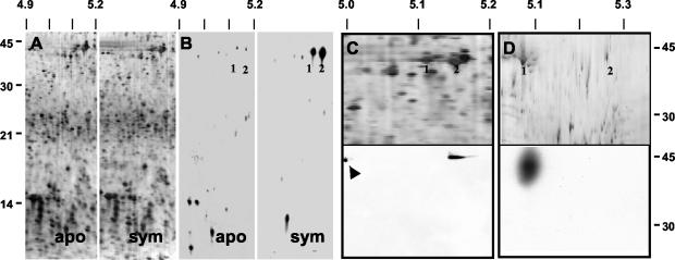
Actin protein was more actively synthesized in 12-h symbiotic light organs. (A) Sectors of silver-stained, 2-D SDS-PAGE gels of soluble proteins (20 μg loaded on each gel) of 12-h aposymbiotic and symbiotic light organs. No reproducible differences in protein profiles could be detected by this method. (B) Autoradiograms of 35S-labeled soluble proteins (20 μg loaded on each gel) of the light organs of aposymbiotic and symbiotic animals. Proteins at a pI and Mr consistent with actin (1, 2) were more actively synthesized in symbiotic animals. (C) Upper panel: sector of a silver-stained, 2-D SDS-PAGE gel of soluble proteins from the light organs of symbiotic animals resolved in the pI range from 4.0 to 7.0. Numbers (1, 2) correspond to those in panel B. Lower panel: immunoblot of the above 2-D gel using antiactin antibodies. Control actin (arrow) was loaded onto the second dimension as a control. (D) Upper panel: sector of a silver-stained, 2-D SDS-PAGE gel of soluble proteins from the light organs of symbiotic animals resolved in the pI range from 4.5 to 5.5. Numbers (1, 2) correspond to those in panel B. Lower panel: immunoblot of the above 2-D gel using antiactin antibodies. Antibody labeling demonstrated that the more acidic of the two proteins (1) at ∼42 kDa was actin. Biotinylated standards were used to determine the Mr. Molecular mass markers for A and B and for C and D appear on the left and right, respectively. pI markers are along the top. apo = aposymbiotic; sym = symbiotic.

